
Table of Contents

Surgical Retina Care
The retina is a thin layer of light-sensitive tissue at the back of the eye that plays a crucial role in vision. When retinal conditions occur, they can affect the clarity of vision and, in some cases, lead to significant permanent vision loss if left untreated.
Surgical retina care focuses on diagnosing and treating retinal conditions that may require specialised procedures and surgeries. At Claris Eye Centre, treatment plans are individualised based on the patient’s specific retinal condition, severity of disease, visual needs and lifestyle, and overall eye health.
Care by Dr Helen Mi Fang
Surgical retina care at Claris Eye Centre is provided by Dr Helen Mi Fang, a dual-fellowship trained ophthalmologist and vitreo-retinal surgeon and medical retinal specialist with extensive subspecialty experience in vitreo-retinal diseases and medical retinal conditions.
Dr Helen manages a wide range of retinal conditions including retinal detachment, macular disorders, retinal tears, myopic retinal conditions, trauma, dislocation of lenses and vitreous haemorrhage. Her practice includes both surgical and procedural treatments for retinal disease, supported by detailed retinal diagnostics and clinical assessment.
Management plans are tailored to the individual’s specific retinal condition, severity of disease and visual needs.
Conditions That May Require Surgical Retina Care
Some retinal conditions may require surgical or procedural treatment to preserve vision or prevent further damage to the retina.
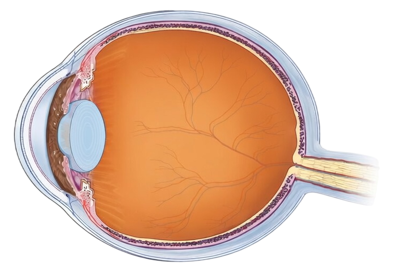
Normal Eye
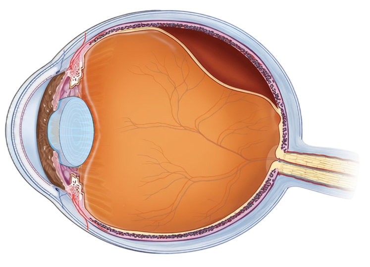
Retinal Detachment
Retinal Detachment
Retinal detachment occurs when the retina separates from the underlying tissue that supports it. This will disrupt normal retinal function and will lead to vision loss if not treated promptly.
Symptoms may include:
- Sudden increase in floaters
- Flashes of light
- A shadow or curtain across the field of vision
- Sudden reduction in vision
Surgical treatment is required to reattach the retina and stabilise vision once detachment occurs. In some cases, the condition may be complex, particularly in patients with significant myopia, trauma, or prior eye surgery.
Normal Eye
Macular Hole
Macular Hole
A macular hole is a defect in the macula, the central part of the retina responsible for detailed and central vision.
Patients may notice:
- Blurred or distorted central vision
- Difficulty reading or recognising faces
- A dark or missing spot in central vision
Surgical treatment (vitrectomy) may be recommended to repair the macular hole, in order to improve or stabilise vision.
Epiretinal Membrane
An epiretinal membrane is a thin layer of scar-like tissue that forms on the surface of the retina centrally over the area called the macula. It can cause the central retinal structures to wrinkle or distort.
Symptoms may include:
- Distorted or wavy vision
- Blurred central vision
- Difficulty reading small print in both distance and near
Depending on the symptoms and visual needs, surgery may be considered if visual function and quality of life is affected.
Vitreomacular Traction
Vitreomacular traction occurs when the vitreous gel remains partially attached to the macula or other parts of the retina, and exerts pulling forces.
This traction may lead to distortion of central vision and blurred vision. There is also a risk of progression to macular hole, and surgery may be considered if symptoms are significant, or if risk is deemed to be high.
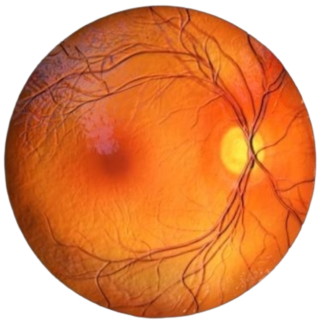
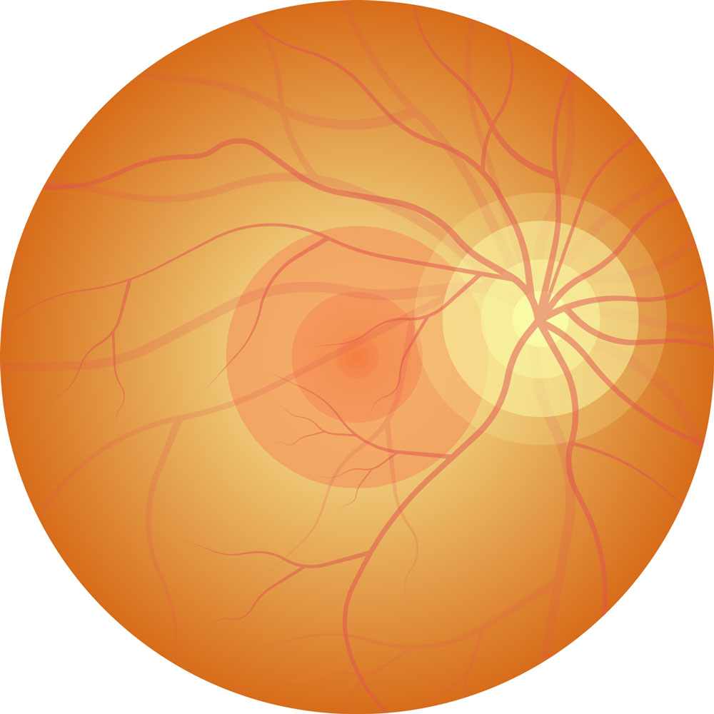
Normal eye retina
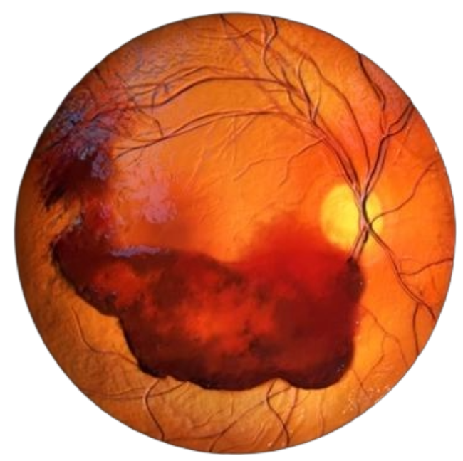
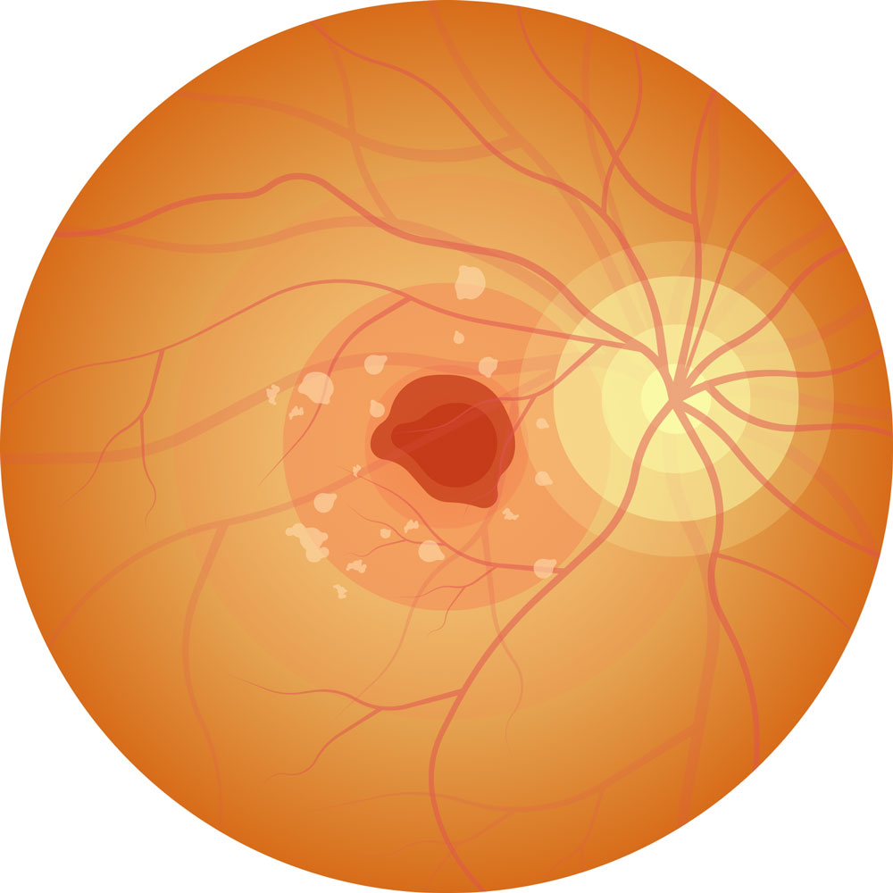
Vitreous haemorrhage
Vitreous Haemorrhage
Vitreous haemorrhage occurs when blood leaks into the vitreous gel inside the eye. This may result in:
- Sudden appearance of floaters
- Dark or red spots in vision
- Blurred or obscured vision
Common causes include diabetic eye disease, retinal tears or trauma. Treatment depends on the underlying cause and severity.
Sub-Macular Haemorrhage
Sub-macular haemorrhage occurs when there is sudden bleeding under the macula, which is the central part of the retina responsible for detailed vision. This can be due to underlying age-related macular degeneration, blood polyps, trauma or other causes.
Symptoms that patients may notice:
- Sudden loss of central vision
- Dark or grey patch centrally
Retinal Disorders Related to Myopia
- Floaters and flashes
- Distorted or wavy vision
- Blurred central vision or peripheral vision
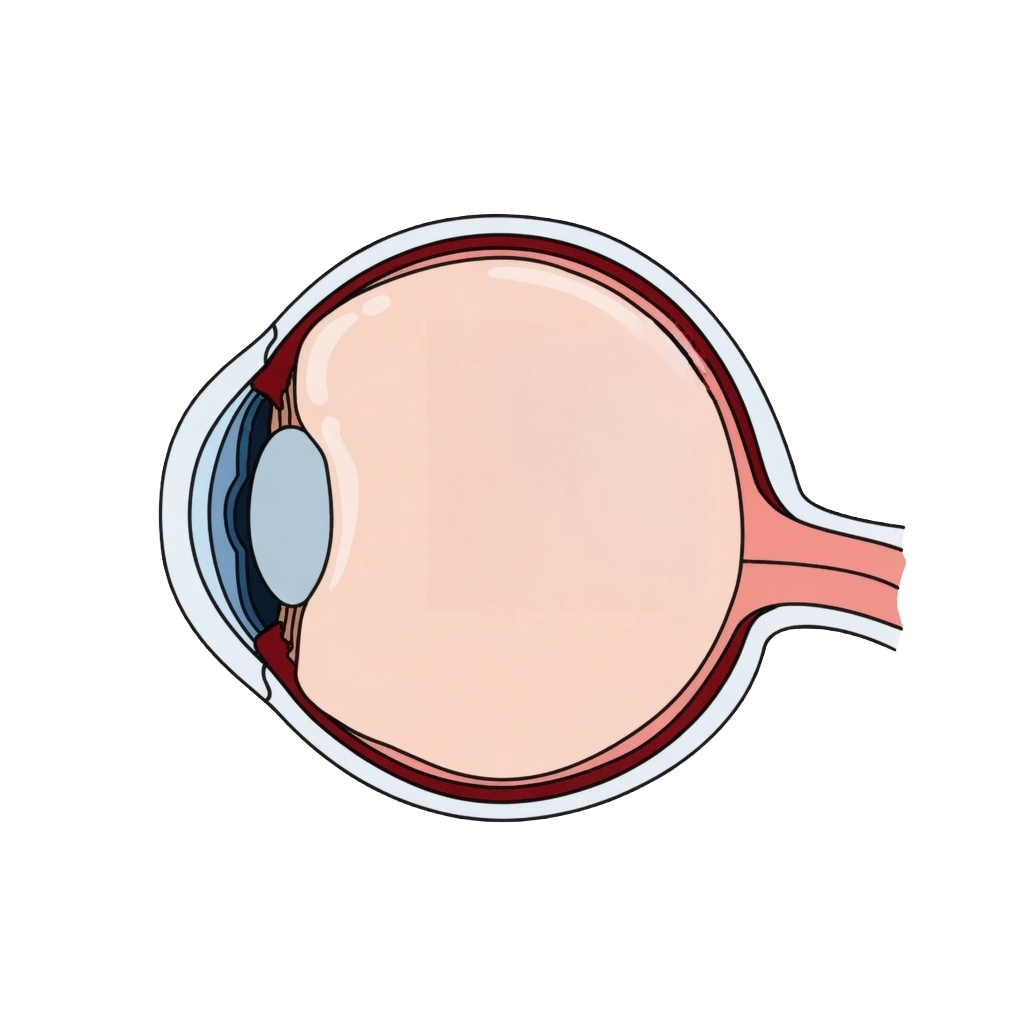
Normal lens
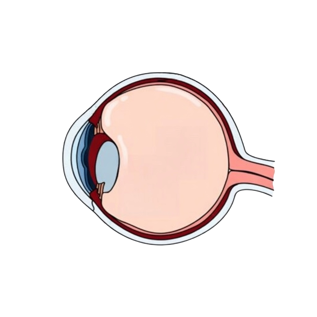
Dislocated lens
Dislocation of Lens
The lens is the structure in the eye that helps with focusing images on the retina. Dislocation of the lens (natural or artificial) can occur due to trauma, extensive rubbing of the eyes, history of eczema, or other medical conditions that predisposes the patient to weakness in the support of the lens.
Patients may notice:
- Sudden blurring of vision
- Inability to focus images
- Double vision
Such patients will require surgery to remove the dislocated lens, and a secondary artificial lens will then be inserted and specially fixed in the eye.
Eye Trauma and Endophthalmitis
Surgical Treatments for Retinal Conditions
Different surgical approaches may be used depending on the type and severity of the retinal condition.
Vitrectomy
Vitrectomy is a microsurgical procedure used to treat a variety of retinal disorders.
During vitrectomy:
- Small “keyhole” wounds are created
- The vitreous gel is removed from the eye
- Specialised instruments are used to repair the area of concern, and laser or cryotherapy may be applied during surgery
- The eye may be filled with gas or silicone oil to support retinal healing
Vitrectomy is used to treat conditions such as retinal detachment, macular holes, epiretinal membranes, vitreomacular traction, vitreous haemorrhage, or severe intraocular infections.
Scleral Buckle
Scleral buckle surgery involves placing a small silicone band around the eye to support the retina and relieve traction.
This technique may be used in certain types of retinal detachment and helps reposition the retina against the eye wall.
Laser Retinopexy
Laser retinopexy is a laser treatment used to seal retinal tears or weak areas in the retina.
The laser creates small burns around the tear, allowing scar tissue to form around the area of concern like a barricade, and secure the retina in place. This helps to reduce the risk of retinal detachment.
Pneumatic Procedures
Pneumatic procedures involve injecting a gas bubble into the eye to help reposition the retina or displace any blood from crucial areas in the retina.
The gas bubble floats and presses against the targeted area of retina, depending on the underlying issue. This technique may be used in selected cases of retinal detachment or submacular haemorrhage.
Symptoms That Should Be Evaluated Promptly
Certain visual symptoms may indicate a serious retinal problem and should be assessed by an eye specialist promptly
These include:
- Sudden increase in floaters
- Flashes of light
- Distortion of central vision
- A dark shadow or curtain across vision
- Sudden decrease in vision
- Obscured central vision
Early evaluation allows for the appropriate treatment to be considered.
Assessment of Retinal Conditions
A comprehensive eye examination is required to diagnose retinal conditions.
This may include:
- Visual acuity testing
- Intraocular pressure testing
- Dilated retinal examination
- Optical coherence tomography (OCT) imaging
- Retinal photography
- Retinal angiography using OCT or intravenous dyes
- Visual field testing
- Ocular ultrasound where required
These tests help determine the cause of symptoms and guide treatment decisions. Further tests may be required based on the individual’s condition.
- Your Retinal Specialist
Dr Helen Mi Fang
Book A Consultation
If you are experiencing sudden visual changes such as flashes, floaters, or distortion of vision, an eye assessment is recommended. Schedule an appointment with Claris Eye Centre for a comprehensive retinal evaluation and discussion of appropriate treatment options.